
5 Shoulder Scenarios That Decide MRI or Ultrasound Use
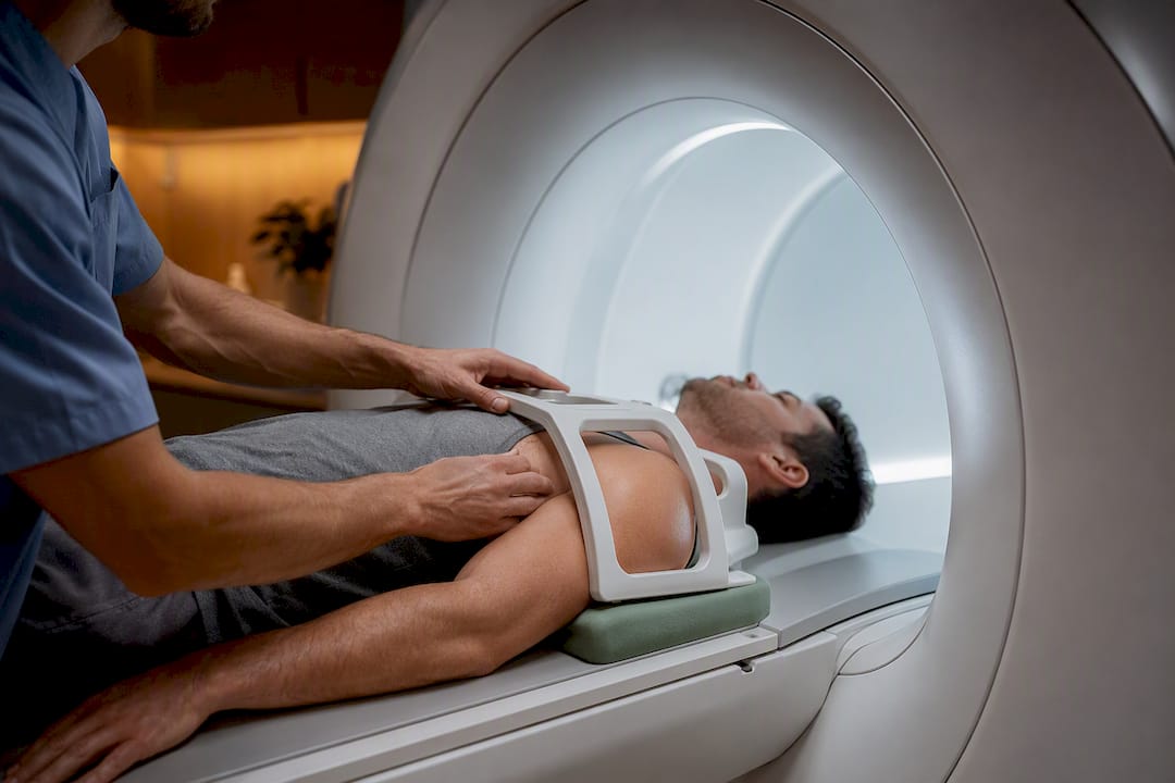
For most suspected rotator cuff tendon problems, a high-quality shoulder ultrasound gives you an accurate, faster, and lower-cost first look, with pooled accuracy comparable to MRI for full-thickness tears. Reserve MRI (or MR arthrography) for cases needing whole-joint detail, surgical planning, labral evaluation, or when the ultrasound result doesn’t match your symptoms.
TL;DR:
Ultrasound is highly accurate for full-thickness rotator cuff tears and offers real-time, dynamic assessment of tendon movement, making it suitable for initial tendon-focused evaluation.
MRI provides comprehensive visualization of deep shoulder structures such as the labrum, cartilage, and bone marrow, and is preferred for complex injuries or surgical planning.
Both tests have similar sensitivity and specificity for full-thickness tears, but partial-thickness tears remain challenging to detect accurately with either modality.
Ultrasound’s operator skill significantly influences accuracy, while MRI interpretation depends on the radiologist’s expertise and equipment quality.
The choice of imaging depends on the suspected injury, patient-specific factors, and whether the goal is quick screening or detailed joint mapping.
Table of Contents
MRI vs Ultrasound Shoulder Imaging: What Each One Actually Shows
Once you understand what each machine is built to see, the choice starts to make sense on its own. Ultrasound and MRI aren’t competing versions of the same test. They’re built to answer different questions, and knowing which question you’re asking changes everything about which one you need.
MRI captures the whole joint in cross-section: bone marrow, cartilage, the labrum (the cartilage ring that stabilizes your shoulder socket), and the rotator cuff tendons, all in reproducible, multi-planar images a radiologist can review later, at leisure, on a screen. A 3.0T (tesla) magnet, the higher-field standard now common at hospital-based centers, tends to resolve small partial tears better than older 1.5T scanners. That resolution matters when a surgeon needs a precise map before opening the joint.
Ultrasound trades that panoramic view for a magnifying glass. High-frequency probes, generally 7.5 MHz or higher, produce remarkably fine detail of tendon fibers, and because the exam happens in real time, the sonographer can move your arm and watch tendons glide (or catch) during motion. That dynamic piece is something MRI cannot replicate on a still image.
The practical differences break down like this:
MRI favors deep or hidden structures: labrum, cartilage, bone marrow edema, and full anatomical mapping before surgery.
Ultrasound favors surface tendon detail, live movement testing, and immediate correlation with the exact spot where you feel pain.
Operator skill matters more for ultrasound. A skilled sonographer can rival MRI accuracy; an inexperienced one may miss a tear MRI would have caught.
MRI accuracy depends on the reader. A radiologist interpreting static images can still miss subtle partial-thickness tears, particularly on older or lower-field equipment.
Ultrasound is typically used first for tendon-focused pain; MRI is typically reserved for surgical candidates or complex, multi-structure injuries.
What the Research Says About Diagnostic Accuracy
The strongest evidence on this question comes from pooled data across many studies, not any single trial. A widely cited meta-analysis found no statistically significant difference between MRI and ultrasound for detecting rotator cuff tears overall, with MR arthrography (MRI performed with a contrast injection into the joint) often edging out both for the highest overall accuracy.
By the numbers: A Cochrane-affiliated systematic review reported pooled sensitivity and specificity for MRI around 0.91/0.89 and for ultrasound around 0.91/0.85 across included studies, meaning both catch the large majority of full-thickness tears and correctly rule out most people without one. The same review found accuracy dropped for partial-thickness tears across all three techniques, MRI, MRA, and ultrasound alike.
That gap on partial tears is the detail most people never hear about. A full-thickness tear, where the tendon is torn all the way through, is relatively easy for any modality to spot. A partial tear, where only part of the tendon’s thickness is damaged, is a much subtler finding, and all three imaging methods struggle with it to some degree.
A few caveats belong here, because the evidence isn’t flawless:
Many of the underlying studies are small, and study quality and patient populations vary widely across the pooled reviews.
Time between imaging and surgery (used as the reference standard in most studies) can introduce error if a tear worsens between the scan and the operating room.
MRA tends to outperform standard MRI and ultrasound specifically for labral tears and some partial-thickness cuff injuries, which is why surgeons often request it before revision surgery.
Here’s what a negative scan actually means in practice: if your ultrasound or MRI comes back clean but your symptoms strongly suggest a rotator cuff problem, that result doesn’t rule it out with certainty, especially for partial tears. It means the next step is usually either a repeat exam with a different technique, an MRA, or a trial of conservative treatment while your provider watches how your shoulder responds.
Which Imaging Test Fits Your Shoulder Symptoms?
Test selection should follow your clinical picture, not the other way around. Here’s how that typically plays out:
Suspected full-thickness supraspinatus tear, no surgery planned yet. Ultrasound first. It’s fast, accurate for this exact scenario, and lets your provider watch your tendon move during the exam.
Suspected full-thickness tear in a surgical candidate. MRI, since a surgeon needs the full anatomical picture, tendon retraction distance, and muscle quality, before planning an operation.
Suspected labral tear, instability, or complex multi-structure injury. Skip ultrasound and go straight to MRI or MRA. Ultrasound cannot reliably visualize the labrum or deep cartilage.
Inconclusive or symptom-mismatched ultrasound result. Move to MRI to resolve the uncertainty, particularly if pain persists or worsens.
Revision surgery planning or complicated prior repair. MRA is often the preferred next step, since scar tissue from previous surgery can confuse both standard MRI and ultrasound.
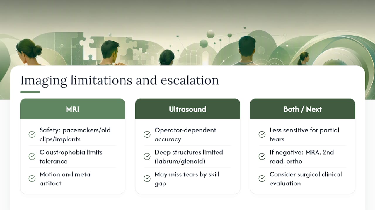
Patient-specific factors can override the usual order entirely. Pacemakers, certain aneurysm clips, and some older metal implants can rule out MRI outright, while severe claustrophobia may make an open MRI or ultrasound the more realistic option regardless of what’s theoretically more accurate. Larger body habitus can also limit MRI image quality in some cases.
Pro Tip: Ask your provider directly whether they’re ordering imaging to confirm a diagnosis or to plan surgery. If it’s the former and you don’t yet have a surgery date, ask whether starting with ultrasound makes sense; it’s often just as informative and gets you an answer in a fraction of the time.
Many clinics run on an ultrasound-first workflow specifically for this reason: it screens efficiently, and MRI gets reserved for the cases that genuinely need its depth.
What to Expect During Your Shoulder Scan
A shoulder ultrasound usually takes 15 to 20 minutes. You sit or lie down while a technician or physician applies gel and moves the probe over your shoulder, often asking you to lift, rotate, or reach your arm so they can watch tendon movement directly and correlate it with where it hurts.
An MRI takes longer, typically 30 to 45 minutes, and requires you to lie still inside the scanner’s tube. If your provider suspects a labral tear or wants a clearer look at cartilage, they may order an MR arthrogram, which involves a radiologist injecting contrast dye into the joint before scanning.
Ultrasound reports are usually written by the same physician or sonographer performing the exam, often noting exact tendon involvement and comparing it to your reported pain point.
MRI reports are written by a radiologist reviewing images afterward, and language like “full-thickness tear” or “high-grade partial tear” signals a clear finding, while “tendinosis” or “indeterminate” suggests a more ambiguous picture worth discussing further.
Ultrasound appointments are often easier to schedule same-week; MRI slots, especially with contrast, may take longer to book depending on your area.
What Shoulder Imaging Costs and Where to Get It
Cost is one of the biggest practical differences between these two tests, and it’s rarely discussed upfront. A U.S. survey of imaging centers found the mean technical cost for a non-contrast shoulder MRI runs about $1,874, with a wide range from roughly $500 to $4,000 depending on facility type. Hospital-based imaging departments tend to charge more than independent, freestanding imaging centers for the identical scan.
Ultrasound is consistently the less expensive option, and because it requires no magnet or dedicated suite, it’s often available at point-of-care in an orthopedic or sports medicine office the same day you’re examined.
A few ways to control your out-of-pocket cost:
Call independent imaging centers directly and ask for self-pay pricing before booking through a hospital system.
Confirm whether the quoted price includes the radiologist’s reading fee separately, since that’s a common surprise charge.
Check with your insurance plan about prior authorization requirements for MRI; ultrasound rarely needs it.
Ask whether ultrasound is available same-day in your provider’s office rather than a separate radiology visit, though availability often depends on business hours since it requires a trained sonographer on-site.
Contraindications, Pitfalls, and When Neither Test Is Enough
MRI has real safety limits that ultrasound doesn’t share. Certain pacemakers, older aneurysm clips, and some metal implants can make MRI unsafe or impossible, and claustrophobia keeps some patients from tolerating the closed tube at all. Motion artifact and metal artifact from prior hardware can also blur MRI images enough to complicate a reading.
Ultrasound’s main limitation is the person holding the probe. An inexperienced sonographer can miss a tear that a skilled one would catch instantly, and deep structures like the labrum and glenoid cartilage are simply outside what sound waves can visualize well.
Both tests underperform on partial-thickness tears compared with full-thickness ones. If your scan comes back negative but your pain and weakness point strongly toward a rotator cuff problem, that’s the moment to ask about MRA, a second opinion read, or a referral to an orthopedic surgeon for direct clinical evaluation.
How Imaging Results Shape Your Rehab Plan
At Contemporaryrehabservices, imaging results almost always change how we structure your care, but not always in the direction people expect. A confirmed partial tear or tendinosis often means conservative rehab first, since research supports ultrasound and MRI both catching enough detail to justify a structured strengthening approach before considering surgery. A full-thickness tear in an active patient may shift that conversation toward surgical consultation alongside our 8 to 16 week rotator cuff rehab program. We often recommend a trial of manual therapy and targeted exercise before imaging when your history and exam already point toward a manageable soft-tissue issue.
Choosing Between MRI and Ultrasound: Our Take
Here’s the honest read: most people asking about shoulder imaging don’t need the most advanced test available; they need the right test for their specific question. Ultrasound answers “is this tendon torn, and how does it move” efficiently and affordably. MRI answers “what does the entire joint look like, and can a surgeon plan around it.” When you book imaging, ask your provider which question they’re actually trying to answer, and don’t be afraid to ask if ultrasound could settle it first. Follow up promptly if a “normal” result doesn’t match how your shoulder feels.
— CRS Wellness
A Path Forward When You’re Focused on Recovery, Not Just Diagnosis
Imaging tells you what’s happening inside your shoulder. It doesn’t rebuild strength, restore range of motion, or get you back to lifting, swimming, or sleeping through the night without pain. That’s where rehabilitation takes over, and it’s where Contemporaryrehabservices comes in.
Whether your scan showed a partial tear, tendinosis, or came back inconclusive, our in-person and telehealth physical therapy sessions build a plan around your actual findings and functional goals, not a generic protocol. Our hands-on approach draws on manual therapy techniques including myofascial work, joint mobilization, and neuromuscular re-education, paired with a phased rotator cuff rehab program designed around your imaging results and recovery timeline. We accept Medicare, Aetna, Cigna, Emblem, and United Healthcare plans, along with direct cash payment, so coverage rarely stands between you and starting care. If you’re near our Albertson or Williston Park locations, book a consultation and bring your imaging report with you; we’ll walk through what it means for your specific recovery plan.
Sources
This article is general information, not a substitute for advice from a qualified doctor. Consult a qualified healthcare professional about your own circumstances before acting on anything here.
FAQ
What can an MRI show that an ultrasound cannot?
MRI can visualize the labrum, joint cartilage, bone marrow, and deep structures that ultrasound’s sound waves cannot penetrate or resolve clearly.
Will a torn rotator cuff show on an ultrasound?
Yes. Ultrasound detects full-thickness rotator cuff tears with accuracy comparable to MRI, though it’s less reliable for smaller partial-thickness tears.
What if my MRI shows nothing but my shoulder still hurts?
A “normal” MRI doesn’t fully rule out a partial tear or tendon inflammation; ask your provider about a second read, an MR arthrogram, or a direct clinical exam, and consider a trial of physical therapy while symptoms persist.
Why do doctors use ultrasound instead of MRI?
Ultrasound is faster, less expensive, has no contraindications, and lets the examiner watch your tendon move in real time and directly correlate findings with your exact pain point.
Can Contemporaryrehabservices help after I get imaging results?
Yes. We build individualized rehab plans, including a structured rotator cuff program, around whatever your MRI or ultrasound findings show, and we accept various insurance plans.
Recommended
Comments