
Ask These Questions First: MRI vs X-ray for Knee Pain

For most knee problems, a weight-bearing X-ray comes first. It quickly rules out fractures, shows arthritis, and checks alignment at a fraction of the cost of an MRI. MRI becomes the right call when a doctor suspects a torn ligament, meniscus, or cartilage damage, or when an X-ray looks normal but your symptoms say otherwise. The exceptions: significant trauma with a suspected hidden fracture, or a young athlete with a classic story for a ligament tear, where MRI or even CT might come sooner.
TL;DR:
Weight-bearing X-rays are the first step for most knee issues, especially for assessing joint space narrowing and osteoarthritis signs.
MRI is preferred for soft tissue injuries such as ligament tears, meniscal damage, or persistent instability that X-rays cannot reveal.
Clinical guidelines recommend starting with X-rays unless trauma or indications of soft tissue injury highly suggest MRI upfront, especially for younger athletes.
MRI costs significantly more and is often delayed due to insurance preauthorization, making X-ray the practical initial test.
For chronic pain and osteoarthritis, X-rays often provide enough information, with MRI reserved for unclear cases or before surgery.
Table of Contents
MRI vs X-Ray Knee Imaging: What Each Test Actually Shows
An X-ray sends a small dose of radiation through your knee and captures the density of bone. It is fast, inexpensive, and remarkably good at answering a narrow set of questions: is something broken, is the joint space narrowing, and is the bone lined up the way it should be.
Doctors often order weight-bearing X-rays rather than the standard version taken lying down, because standing loads the joint the way it behaves in real life. A knee with early osteoarthritis can look deceptively normal on a non-weight-bearing film. Put your body weight through it and the joint space narrows, revealing wear that would otherwise stay hidden.
Common findings on a knee X-ray include:
Fractures, including hairline breaks along the tibial plateau or patella
Osteophytes, the bony spurs that signal osteoarthritis
Joint-space narrowing from cartilage loss
Malalignment, such as bowlegged or knock-kneed patterns
Loose bodies, small fragments of bone or calcified cartilage floating in the joint
Where X-rays fall short is soft tissue. A radiograph cannot see your meniscus, your ACL, or the cartilage surface itself. It also can’t detect bone marrow edema, the swelling inside bone that often shows up after a stress injury long before a fracture line appears. If your knee looks fine on X-ray but keeps giving out or locking, that gap is exactly why a clinician moves to a different tool.
MRI Knee Benefits: Seeing the Soft Tissue X-Rays Miss
Magnetic resonance imaging uses magnetic fields rather than radiation, and it’s built for exactly the tissue X-rays overlook. MRI is considered the preferred test for evaluating internal derangement of the knee, the umbrella term for tears and damage inside the joint that a plain film simply cannot capture.
An MRI knee scan typically reveals:
ACL and PCL tears, including partial versus complete disruption
Collateral ligament injuries on the inner or outer knee
Meniscal tears and their exact location and pattern
Cartilage lesions on the joint surface
Bone marrow edema, often a sign of bruising or early stress injury
Synovitis, or inflammation of the joint lining
Doctors typically reach for MRI when a patient reports instability, a sense of the knee giving way, mechanical locking, or symptoms that persist despite a clean X-ray. It’s also standard before knee surgery, since a surgeon needs to know exactly what they’re working with before making an incision.
One catch worth knowing: MRI can find things that don’t matter clinically. Many meniscal tears and cartilage changes that show up on MRI in middle-aged and older adults may exist without causing any symptoms. An MRI knee report full of abnormal-sounding findings doesn’t automatically mean surgery is the answer. It means the images need to be read alongside your actual symptoms and a physical exam, not in isolation.
How Do Doctors Decide Between X-Ray and MRI for a Knee?
Clinical guidelines exist precisely so this decision doesn’t come down to guesswork. The ACR Appropriateness Criteria recommend radiographs as the initial imaging study for the large majority of acute and chronic knee complaints, reserving MRI for cases where soft-tissue injury is suspected or an X-ray leaves the picture incomplete. A 2026 update to that guidance reinforces radiographs as the default first step for chronic knee pain, while spelling out the narrower scenarios where MRI is appropriate right away.
In practice, the decision tends to follow a short sequence:
Take a history and do a hands-on exam. Swelling, range of motion, and specific stress tests (checking ligament stability, for instance) narrow the possibilities before any scan is ordered.
Apply clinical rules for trauma. Tools like the Ottawa knee rules help determine whether a fracture is likely enough to justify an X-ray at all after an acute injury.
Order imaging based on age and mechanism. A twisting injury in a 22-year-old soccer player points toward possible ligament or meniscal damage. Knee pain that’s built gradually in a 65-year-old points toward osteoarthritis and a weight-bearing X-ray.
Escalate to MRI only when the picture stays unclear, or when surgery is genuinely on the table.
What Do X-Ray and MRI Cost, and What Are the Risks?
Cost is usually the first practical difference patients notice. X-rays are inexpensive and rarely require prior insurance approval. MRI runs considerably higher and frequently needs preauthorization, which can add days to the process before you even get scheduled. If you’re weighing out-of-pocket costs for any advanced scan, resources like this breakdown of DEXA scan costs give a useful sense of how imaging pricing works outside insurance networks, even though DEXA and MRI serve different purposes.
Timing differs sharply too. An X-ray takes just minutes. An MRI scan itself runs 20 to 45 minutes, and that’s before factoring in the wait to get an appointment.
X-rays use a small dose of ionizing radiation, low enough that it’s rarely a deciding factor.
MRI uses no radiation, but carries its own limitations: certain implants and pacemakers can rule it out entirely.
Claustrophobia is a real barrier for a fair number of patients facing a standard closed-bore MRI machine.
Contrast dye, when used, carries its own small set of risks and requires a kidney-function check beforehand.
Which Imaging Fits Which Knee Problem?
Matching the injury to the test avoids wasted time and unnecessary costs. Here’s how the decision usually breaks down by scenario.
Fracture or major acute trauma: X-ray first, always. If the X-ray is negative but suspicion stays high (a fall from height, a direct blow with severe pain), a CT scan may follow to catch fractures a plain film misses.
Sudden instability, locking, or a suspected ACL or meniscus tear: MRI, but only after a focused physical exam has already pointed toward a soft-tissue injury rather than a fracture.
Chronic knee pain or suspected osteoarthritis: Weight-bearing X-rays first. A comparison of radiographs, MRI, and arthroscopy found only moderate agreement between MRI and surgical findings for arthritis, and radiographs frequently gave clinicians enough information on their own. MRI only enters the picture if pain is far worse than the X-ray suggests, or surgery is being planned.
Persistent swelling, suspected bone lesion, infection, or tumor: MRI is usually indicated, since these problems live in soft tissue or bone marrow that a plain film can’t resolve.
What Should You Ask Before Getting an Imaging Test?
Getting the right scan starts with the right conversation. A few direct questions can save you a second appointment and an unnecessary bill.
Ask whether the result will actually change your treatment. If the plan is physical therapy either way, an MRI may not move the needle.
Ask whether you should get a weight-bearing X-ray first, particularly if arthritis or chronic pain is the concern.
Ask about insurance coverage and preauthorization for MRI before you schedule it, since surprise costs are common.
Prepare properly. Remove metal jewelry for either scan, mention any implants or pacemakers before an MRI, and flag claustrophobia so staff can plan accordingly.
Pro Tip: Bring a written timeline of your symptoms, what makes them worse, and what you’ve already tried. A clear history often narrows the imaging decision faster than the scan itself.
Once results come back, resist the urge to treat the report as a verdict on its own. Imaging works best when read alongside your exam and symptoms, not as a standalone diagnosis.
How CRS Wellness Approaches Imaging Decisions
CRS Wellness generally favors an exam-first approach. When osteoarthritis looks likely, a weight-bearing X-ray usually makes sense before anything more advanced. Many patients with mechanical knee pain improve with a focused trial of physical therapy before advanced imaging is even necessary. When symptoms suggest something imaging alone can clarify, the clinic helps patients understand what a referral for MRI would actually add. If you’re unsure whether your knee pain needs a scan at all, resources like signs you may need physical therapy are a reasonable place to start.
Why the Imaging-First Instinct Often Backfires
The instinct to request an MRI immediately is understandable. It feels thorough. But the evidence tells a more nuanced story. Clinical assessment alone is often sufficient to diagnose osteoarthritis, and ordering advanced imaging before a proper exam frequently uncovers findings that were never causing symptoms in the first place. That leads to more anxiety, more follow-up visits, and sometimes procedures that don’t actually address the pain.
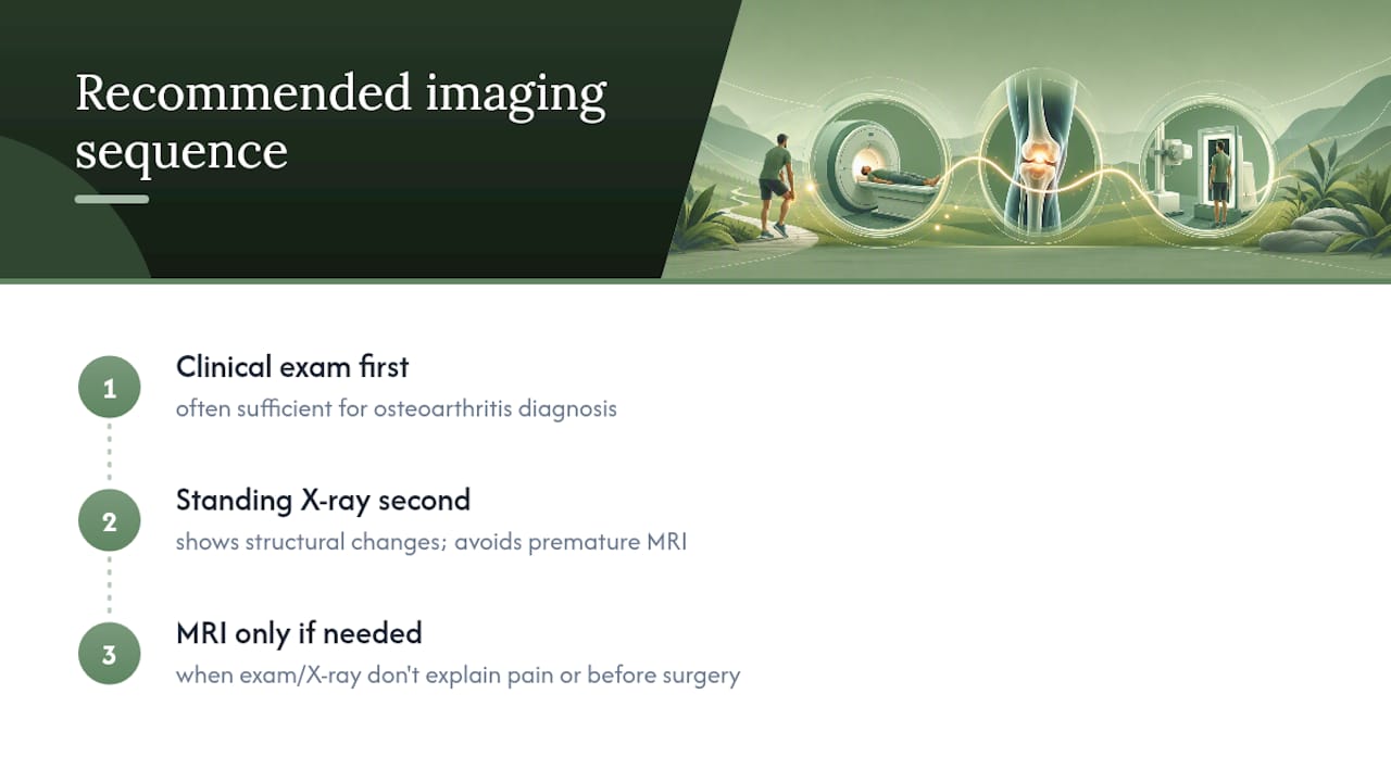
The conventional advice to “just get an MRI to be safe” undersells how much a skilled exam and a standing X-ray can reveal. What the research actually supports is sequencing: exam first, X-ray second, MRI reserved for when the story doesn’t add up or surgery is genuinely being considered. Patients who prioritize that order tend to avoid unnecessary scans, unnecessary costs, and the false alarm of an incidental finding that never needed treatment.
If there’s one thing worth changing in how people approach knee pain, it’s this: ask what a test will change before you agree to it. That single question cuts through most of the confusion around imaging.
— CRS Wellness
A Practical Next Step Before You Book Any Scan
Before you schedule an X-ray or an MRI, it’s worth finding out whether your knee pain even needs one yet. CRS Wellness offers physical therapy sessions and accepts a variety of insurance plans and payment options, so cost doesn’t have to be the reason you skip an evaluation.

A hands-on assessment can often tell you whether your symptoms match a pattern that responds well to conservative care, or whether imaging genuinely makes sense as a next step. For knees where arthritis or wear-and-tear pain is the likely culprit, CRS Wellness’s approach to knee osteoarthritis physical therapy starts with understanding what’s actually driving the pain before recommending any scan. If you’re in Albertson or the surrounding area, you can book an evaluation at the Albertson location and get a clear answer on your next step.
This article is general information, not a substitute for advice from a qualified doctor. Consult a qualified healthcare professional about your own circumstances before acting on anything here.
Sources
Recommended
Comments